


With serious internal diseases, poor nutrition, as well as with age, nail growth slows down, its structure undergoes changes. Only a doctor can accurately determine the cause of the violation based on test results and microscopic examinations.
But to get an idea of what happens to the nails on the feet or hands, you can use the photo with fungal diseases of different types.
Causes of nail deformity
Molds, yeast-like fungi and dermatophytic fungi cause infectious nail diseases (onychomycosis), which have similar symptoms.
All types of fingernails or fingernails deform the nail plate, change the transparency, shine, color, this variety is shown in the photos shown.
Changes in the nail occur not only with onychomycosis, but also with injuries, chronic toenails (inflammation of the nail fold), psoriasis, hand eczema, dermatitis. Before concluding that there is a fungal infection, you should consider all the possible options.
Signs of fungal infection
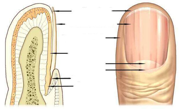
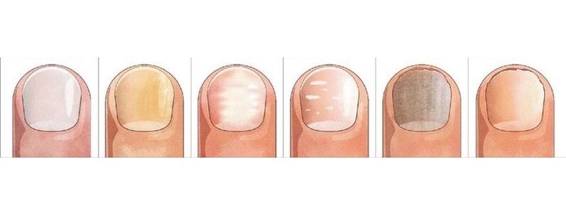
The most informative signs of fungal infection are changes in the color of the nail plate, the presence of nail detachment, superficial changes - transverse, longitudinal grooves in the nail plate, depressive points, thickening, destruction of the nails.
The pink color of a healthy nail is determined by the transparency of the nail plate and the blood vessels that are visible through it. With onychomycosis, the nail loses its transparency, the color becomes brown, yellow, less often green, black.
Candida fungi and dermatophytes cause onycholysis - the separation of the affected part of the nail. When infected with dermatophytes, onyolysis is observed from the distal tip of the nail and when infected with Candida, the nail lags behind the nail bed at the base, in the area of the crescent.A symptom of an authentic fungus may be inflammation of the lateral perivascular ridges - the paronychia. This disease has bacterial forms caused by streptococci and staphylococci, as well as non-infectious - eczema, psoriasis, systemic vasculitis.
When nails are affected by the fungus Trichophyton rubrum, the plaque is affected, as you can see in the photo, the roller is not affected by the infection. The plaque becomes yellowish, thickens intensely, the accumulated fungal masses are well visible below it.
Nail fungus due to dermatophyte infection
In 95% of all nail fungus cases, the disease is caused by dermatophytic Trichophyton rubrum and Trichophyton mentagrophytes.
Trichophyton rubrum infection
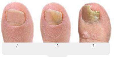
Onychomycosis begins when the fungus penetrates under the nail plate from the free edge. Fungal infection is indicated by the appearance of a yellowish spot, an abnormal, collapsing surface of the peripheral (distal) tip of the nail in the area of the spot.distal-lateral formTrichophyton rubrum dermatophyte infection is common. In the photo, you can see that the stain caused by the introduction of the fungus is located along the lateral peripulmonary fold of the nail.
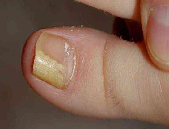
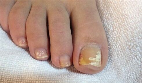
The fungus Trichophyton rubrum typically affects the big toes, causing hyperkeratosis - a buildup of fungus between the nail plate and the nail base, which looks like a loose yellowish photo.
At this stage, the fungus occupies an insignificant part of the nail, as shown in the photo, and with the help of topical treatment it is possible to treat the person starting onychomycosis.
Without treatment, the stain grows, gradually affects the entire tip of the nail and then moves to the half moon. In the photo, the nail fungus looks like yellowish stripes that are directed towards the growth zone of the nail plate.
Withthe peripheral form of nail fungus, often found on the big toes, a yellowish spot of infection appears at the periphery of the nail, in its central part, as can be seen inPhoto.
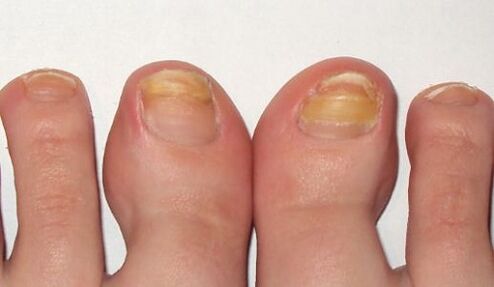
In the advanced stage of the fungus on the feet, several nails are affected, as in the photo, and the treatment is no longer limited to topical treatments and pills. In addition to antifungal drugs, the nail is cleaned with a material to remove all or part of the nail plate.
Long-term treatment using all known antifungal agents and treatments should be performed on the foot, caused by Trichophyton rubrum, with hyperkeratosis, as shown in the photo.Fungal infection with total nail damage spreads to the entire area of the nail plate, the nail is completely destroyed.
Infection with another dermatophyte, Trichophyton mentagrophytes, can also lead to a total fungal infection of the nail. Trichophyton Mental InfectionWith complete defeat of the nail with the fungus Trichophyton mentagrophytes, the nail plate is deformed, the photo shows that it thickens, changes its structure, collapses, yellowish spots appear on its entire surface.
Infection of the nail with this dermatophyte usually causes superficial white onychomycosis of the big toe, less often of the small toe.
This fungus does not appear on the fingernails, often causes transgenic dermatitis on the feet, as in the photo, and requires simultaneous treatment of the skin of the feet and nails.
A symptom of a fungal infection of the nails, usually on the feet, are white spots of various sizes, as in the photo, reminiscent of leukonia - a disease of the nail plate itself.
But unlike leukemia, in which white spots are caused by air bubbles in the nail bed, white spots in a fungal infection are the result of Trichophyton mentagrophytes activity.
Rarely, superficial white onychomycosis is caused by molds. In AIDS, the causative agent of this type of fungus may be Trichophyton rubrum and it affects the nails on both feet and hands.
Changes in the nail due to Candida infection
The fungus usually occurs in women, affecting the nails on the working hand, which is most often in contact with water.
For true onychomycosis, the proximal form of infection is characteristic, in which the fungus first affects the nail fold of the nail base, and then penetrates the growth zone and the nail base. It then gradually moves along the nail from the base to the tip, recording an ever-increasing area of the nail plate.
The causative agent of the disease in candidal onychomycosis is Candida albicans. This fungus invades the nails and toenails, spreading from the half moon zone to the base of the nail plate, to the free edge, as shown in the photo.
A sign of Candida nail infectionalbicans is the inflammation of the nail fold (paronychia), the separation of the skin from the nail plate, the pain, the rejection of a nailbacterial infection.
Candida albicans can penetrate the nail from its free edge. In this case, they are talking about the peripheral form of the infection, which is usually combined with cutaneous candidiasis.
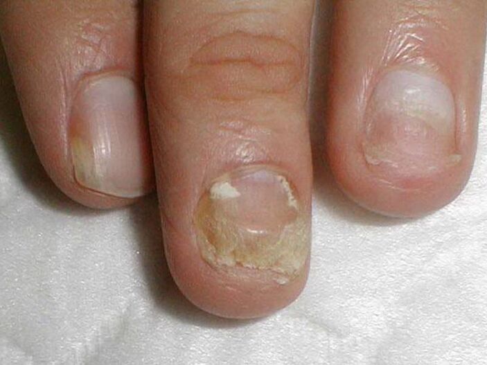
Treatment of candida on the nails of the hands and feet with damage to more than half of the area of the nail plate, as in the photo, includes not only the fight against onychomycosis, but also measures to reduce itcandida activity in their natural storage reservoirs - the intestines, the oral cavity, the mucous membranes of the genitals. . .
Infection by molds
Molds cause fungus much less often than Candida or dermatophytes. The main symptom of mold nail infection is, as you can see in the photo, the change in color of the nail plate to blue, black, greenish.
The marks of the nail mold can be dark spots, dots on the nail plate or, as in the photo, a black longitudinal stripe.
Preparations against fungi
Antifungal agents with fluconazole, ketoconazole, terbinafine, itraconazole, grisofulvin are used to treat nail fungus caused by dermatophytes, as in this photo.Terbinafine antifungal agents are effective for dermatophytic infections.
Antifungal agents with voriconazole are very active against dermatophytes.Usedandto treat nail moldon feet, hands andagainst candida dough. The spectrum of action includes molds such as Aspergillum, Fusarium, Penicillium.
Itraconazole-based preparations treat molds.
Fungal-like nail diseases
A grayish tingesometimes appears on the nailwith eczema. In this case, the nail plate can be removed from the nail bed, which is observed with fungus.
Externally very similar to onychomycosismanifestations of psoriasis. With this disease, not only doeschange color, but it alsothickens the nail plate.
Depression notes on its surface, the separation of the nail plate from the nail layer is noted. But there are differences from the fungus: in psoriasis, the detached and healthy parts of the nail are separated by a pink, yellowish band over time.
Blue colorgets the nailwith pseudomonas nail infection. Frequent mechanical friction of the nail fold causes the appearance of surface grooves, wavy nail.
White spots on whiteness, the appearance of whichis associated with metabolic disorders, can also be perceived as a superficial white fungus with a large area of the spot.
Changes in color, the shape of the nail that causes injury. The big toes are at greater risk. Injured nail, as well as fungus, thickens and darkens

The difference between injury and fungus is that changes during injury occur only in the injured finger, the nails on the other fingers remain unchanged, they are not infected by the diseased finger, as in the diseased finger.
The consequence of the wound may be a partial separation of the nail from the nail layer, the formation of a cavity, which, under adverse conditions, quickly colonizes fungi.
The nail plate can be separated from the nail layer under the influence of light (photoniolysis), with iron deficiency anemia, hormonal diseases. Separation, nail loss occurs with erythematous lichen, blistering dermatitis, nail trauma.
But you can finally make sure the conclusion is correct and start treatment, you can only after seeking help from a dermatologist - a dermatologist or a mycologist - a doctor treating fungal diseases.